IT-11-P-1723 Direct Observation of Magnetic Domain Walls by Lens-Less Foucault Imaging (LLFI)
Lorentz microscopy, categorized as having Fresnel and Foucault modes, is as a practical technique for observing magnetic properties by using a transmission electron microscope (TEM). The Fresnel mode is more popular because it does not require any special equipment in its electron optical system. The Foucault mode, however, requires a magnetic-field shielding lens and an off-center objective aperture. Although this mode can visualize magnetic domains under in-focus conditions, it has been considered impractical. Recently, however, a novel Foucault imaging method, named lens-less Foucault imaging (LLFI) [1], was developed for conventional TEM without any special equipment.
Figures 1(a), (b) and (c) show the optical systems of different modes of LLFI in a 300-kV field emission TEM (HF-3300; Hitachi High-Technologies Corp.). Figures 1(a) and (b) are for observing Foucault images and (c) is for small angle electron diffraction (SAED) pattern. The objective lens was switched off and the electron beam was focused with a condenser lens to the crossover on the selected area (SA) aperture plane. The SA aperture was used as an angular aperture selecting for appropriate Foucault images, and the focal length of the magnifying lens was changed in order to observe the Foucault images and diffractions. The irradiated area on the specimen was set by selecting an appropriate diameter for the condenser aperture. Figure 1(d) is an example of SAED of a 90° ferromagnetic domain structure of La0.75Sr0.25MnO3 (LSMO). The observation was done with a camera length of 150 m. White circles in the lower panel of Fig. 1(d) indicate the diameters and positions of the SA aperture for the LLFI observations and lowercase letters of individual circles correspond to Foucault images in Figs. 1(e) – (l).
Figures 1(e) – (h) show Foucault images of each dispersed deflection spot (see in Fig. 1(d)). The magnetization structure among the 90° domains is directly visualized. Figures 1(i) – (l) show Foucault images of the magnetic domain walls in the same area as Figures 1(e) – (h). Figure 1(i) shows 90°/180° domain walls imaged with four streaks including the optical axis by using an SA aperture 5 μm in diameter. Figure 1(j) is an image of 180° domain walls made using an SA aperture 3 μm in diameter. Figure 1(k) and (l) are 90° domain walls imaged with individual streaks from the upper and lower positions on the optical axis.
The LLFI method is advantageous for observing not only magnetic domains and domain walls but also SAEDs less than 2×10⁻⁵ rad. In combination with high-angular-resolution imaging, it will open the way to developing new applications in Lorentz microscopy.
References:
[1] Y. Taniguchi et al., Appl. Phys. Lett., 101, 093101 (2012).
The authors would like to thank Prof. S. Mori of Osaka Prefecture University for supplying the LSMO specimens and valuable discussion about the materials.
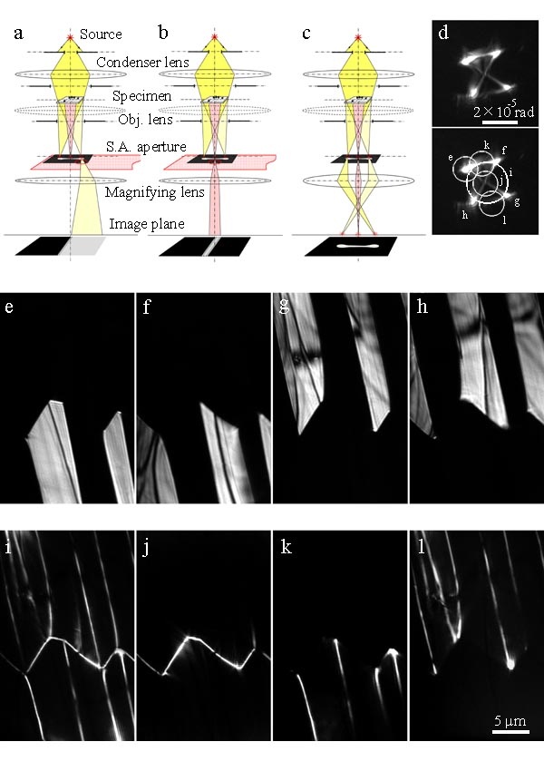
Fig. 1: (a) Optical system for domain observation, (b) for domain-wall, (c) for small-angle electron diffraction, (d) SAED from 90°/180° domain structure of LSMO, (e)–(h) Foucault images of 90°/180° domains from each single deflection spot, (i) –(l) Foucault images of domain walls from streaks, (j) 180° domain walls, (k) and (l) 90° domain walls. |