IT-8-O-1703 Electron Pulse Properties and PINEM Aberrations in Ultrafast Transmission Electron Microscopy
In ultrafast transmission electron microscopy (UTEM), extension of the static imaging and analytical capabilities of transmission electron microscopy to the ultrafast temporal domain relevant for many atomic and nanoscale processes allows for visualization of non-equilibrium structural phenomena. As in pump/probe spectroscopic techniques, the operating principle of UTEM requires – at some point during the experiment – temporal overlap of the femtosecond photon and electron pulses at the specimen. At time zero (i.e., precise overlap of the pulses), significant photon absorption by the freely-propagating electrons and population of virtual states occurs, and peaks occurring at integer multiples of the photon energy can be observed to the gain-side of the zero-loss peak in electron-energy spectra. Because this process, called photon-induced near-field electron microscopy (PINEM), is observed when the pulses are overlapped in space and time at the specimen, proposals for using this phenomenon to measure the total UTEM response function and the electron pulse shape and duration emerged during the initial experimental observations.
In this talk, we will discuss considerations for isolating the inherent artifacts of the highly non-linear near-field interactions from the actual pulse characteristics. Using theory developed to describe these interactions, we will discuss how temporal cross-sections of peaks in the electron-energy spectra corresponding to high-order transitions are expected to exhibit the true temporal behavior of the electron pulses. In general, the exceedingly small portion of the pump laser pulse capable of initiating such transitions results in the temporal widths converging to the electron packet duration. Additionally, population of quantized virtual states occurring for an electron beam focused on the edge of a nanostructure suggest that the resulting energy distribution may produce well-defined chromatic aberrations in images arising from the velocity-dependence of magnetic lens focusing (Fig. 1). As such, we will discuss the prospect for detecting such phenomena and its potential as a means of determining the UTEM instrument response without the need for a spectrometer. Appropriate interpretation of observed spectroscopic and image features should in principle enable systematic temporal and spatial deconvolution allowing for a more accurate depiction of intrinsic ultrafast dynamics, especially the critical initial excitation rising edge which, as advances continue, will drop below 50 fs.
This work is supported by 3M, and acknowledgment is made to the Donors of the American Chemical Society Petroleum Research Fund.
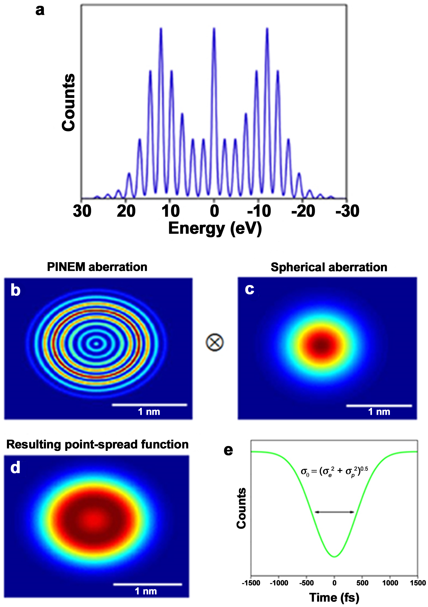
Fig. 1: (a) PINEM energy spectrum. Real-space annular point-spread functions due to (b) quantized chromatic aberration (i.e., PINEM aberration) and (c) spherical aberration. Convolution results in the point-spread function shown in (d). (e) Temporal variation of Fourier intensity for a discrete frequency arising from the convoluted aberrations. |