IT-14-P-1654 Investigating electrical charged samples by scanning probe microscopy: the influence to atomic force microscopy and magnetic force microscopy image artifacts.
The electric state of a surface is of great importance for its chemical and physical properties, e.g. bounding of molecules, charge transfer or dielectric properties. Various scanning probe microscopy techniques based on atomic force microscopy (AFM) map electric surface potentials, electrostatic forces as well as the topography to distinguish electrostatic from van der Waals forces. However, the signal acquired from van der Waals forces can also show artifacts arising from electrostatic forces. Even so such effects are of great importance to interpret image obtained from charged surfaces, the effect is not discussed in detail in literature.
In this work, AFM artifacts resulting from electrical charged surfaces are investigated. In a detailed and systematic study, the influence of an electric field gradient above sample to topography, phase and magnetic force microscopy (MFM) images is investigated. Images were acquired with a commercial AFM using a lithographical patterned Kelvin force microscopy (KFM) calibration sample (Fig. 1). Our results show that electrical charges give rise to a signature in topography (Fig. 2) and phase signal. In order to minimize these artifacts, they are studied in regard of various acquisition parameters. We find that either using a low relative set point or high free vibration amplitude during images acquisition reduces the influence to the AFM measurements. Both approaches can sufficiently negate the effect by increasing the tip/sample interaction (either by getting the tip closer to the sample surface or by larger tip vibration amplitudes). As a trade off to these approaches, the sensitivity to topography features is reduced.
Finally, commercial metalized MFM cantilevers are studied in regard their sensitivity to electrical charge present on the sample surface. We observe the appearance of a MFM contrast for the non-magnetic KFM test structure (Fig. 3) for such conditions. The electrical charges give rise to a MFM signature indistinguishable from a magnetic signature exhibiting a strong correlation to KFM images obtained from our sample. The results indicate that great care has to be taken in the interpretation of topographic, phase contrast or magnetic images, when electrical fields are present on the sample surface.
This research was financially supported by Ministério da Ciência, Tecnologia e Inovação (MCTI) - Brazil.
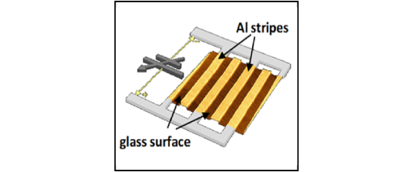
Fig. 1: Illustration of KFM test sample consisting of interdigitated Al stripes on a glass substrate. |
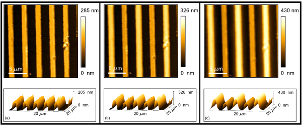
Fig. 2: AFM topography images (a) 0V, (b) 5V and (c) 10V applied between the finger pairs. |

Fig. 3: (a)Topographic image and “MFM” phase shift for our KFM test sample (b) 0 V and (c)5V applied between the finger pairs. |
|