IT-2-P-1525 Cs CORRECTED ATOMIC RESOLUTION TEM IMAGES OF THE HUMAN TOOTH ENAMEL CRYSTALS
Aberration-corrected HR-STEM and HAADF images of the human-tooth-enamel crystallites are presented. These spatial and energy resolutions images have allowed to get information on the physical meaning of the central dark line (CDL) defect which leads to the anisotropic dissolution of the crystals.
Human tooth enamel is composed in 95% of hydroxyapatite crystals (HAP, Ca10(PO4)6(OH)2) which are elongated-plate-like of 30 to 60 nm wide and 100 to 200 nm long, approximately [1]. They are organized in microns-sized structures named “rods” or “prisms” that go from the enamel–dentin junction to the enamel surface (figure 1). The chemical analysis in the micron range of enamel by different analytical techniques, mainly EDS spectroscopy, has indicated the existence of trace elements. Thus, carbonated hydroxyapatite (c-HAP) with Na, Mg, Cl, as trace elements, has been stabled for these crystals [2].
When observed with the Transmission Electron Microscope (TEM), the enamel crystallites show a structural defect of 1 to 1.5 nm width in their central region approximately, the Central Dark Line (CDL) (figure 2), whose structure and role in the enamel structure itself is unknown yet [3, 4].
Several studies have shown that the CDL favors their anisotropic dissolution [4, 5]. During the carious process, for example, the enamel crystals are destroyed in a systematic fashion: first a series of hexagonal holes aligned along the [11-20] are observed, then the holes develop anistropically along the [0001] direction and cross the whole crystals [4, 5].
Enamel crystals are electron beam sensitive, other important parameter against the HRTEM observation (figure 3). Therefore, the use of low electron doses is critical during the study of the CDL. Therefore aberration corrected HR-STEM is the appropriated equipment for carrying out the chemical and structural analyses of the enamel crystallites.
Human tooth enamel samples were obtained from permanent non-carious human molar teeth, extracted for orthodontic or periodontal reasons. Samples were prepared in the FIB-FEI QUANTA 200 3D equipment using the two beams system.
JRG thanks to DGAPA-UNAM (contract IN106713), CONACYT and PASPA-DGAPA-UNAM for sabbatical support.
References
1. R.Z Le Geros, Calcium Phosphates in Oral Biology and Medicine ed H M Myers (San Francisco, CA: Karger). 1991.
2. G.E. Tiznado-Orozco et al., J. Phys. D: Appl. Phys. 42 (2009).
3. J. Reyes-Gasga et al., J. Mater. Sci. Mater. Med. 19, 877-882 (2008).
4. E. Brès et al., Journal de Physique, 51, C1-97-102, (1990).
5. E. Brès et al., Ultramicroscopy, 12, 367-372 (1984).
This research has received funding from the European Union Seventh Framework Programme under Grant Agreement 312483-ESTEEM2 (Integrated Infrastructure Initiative–I3).
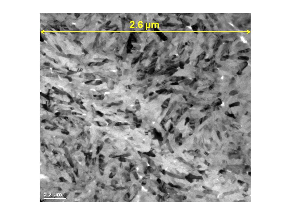
Fig. 1: TEM image of human tooth enamel crystals inside the micron-sized structure named “prism”. |
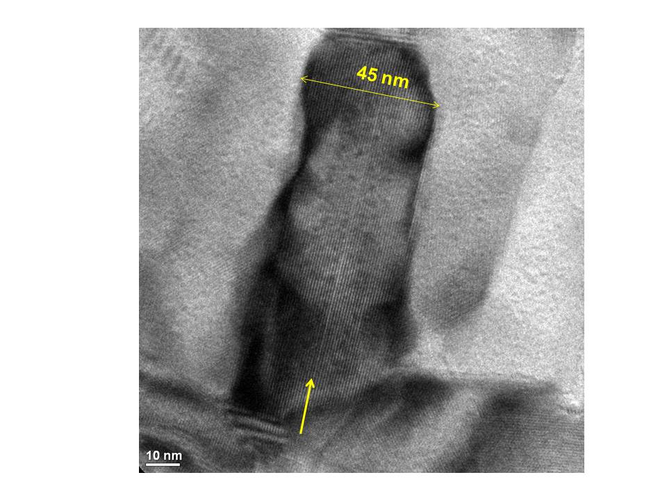
Fig. 2: Magnification of one of the human tooth enamel crystals shown in figure 1. The row indicates the presence of the “central dark line”. |
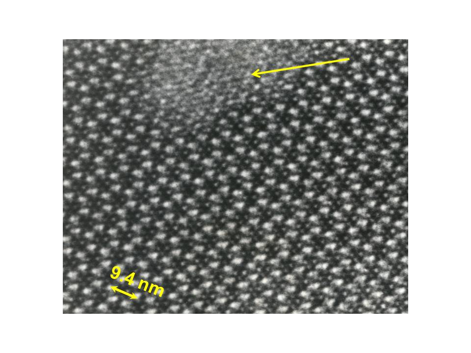
Fig. 3: HRTEM image of a human tooth enamel crystals aligned along the [0001] direction. The arrow indicates the electron beam damage. |
|