IT-13-P-1477 Development of Cryo-FIB technique for the structural characterization of liquid samples
It is important to observe and characterize the three-dimensional distribution of materials in liquid samples such as; cosmetics, functional paint, catalysts and similar products. Furthermore the requirement for investigating the structure inside of liquids at a microscopic level such as the interface of a dispersoid and dispersant has increased. In order to meet these requirements, we have developed a fully compatible cryo transfer holder for FIB and (S)TEM systems. Another area of development for this holder centers on controlling the temperature of the specimen during either the fabrication or observation and which makes it possible to transport a specimen in the frozen condition. This holder can be cooled to 100K by liquid N2 for observation or fabrication of the sample in a controlled thermal state. To demonstrate the capabilities of this holder a liquid foundation sample was investigated. The liquid foundation was first plunge frozen on a specimen stub and transferred to the holder. The holder containing the frozen foundation sample was then placed into a Hitachi NB5000 FIB-SEM for FIB fabrication.
As a result 100nm thin lamella was produced in the FIB and we were able to observe the structure of the microscopic dispersoid, and its distribution as first viewed in the NB5000 and then a HD2700 shown in Figure1.
Figure 2 shows the EDX maps of the thin foil specimen (Thickness: approximately 100nm) of the frozen foundation. In this result it is possible to clearly see each dispersoid, such as the Fe needle crystals and the granular Ti containing crystals. The results confirm the low temperature and high stability performance of this cryo-transfer holder.
In conjunction to this cryo holder a modified method for the micro-sampling technique which allows a micrometer sized sample to be taken from a millimetre sized specimen is required. For this a new mechanical cryo-probe was developed for sample extraction. This mechanical cryo probe is able to be cooled to 120K.
Figure 3a show the cross-section SEM images of a frozen facial foundation liquid which was lifted out by using a room temperature probe needle. Some hollows appeared inside the micro sample due to the water sublimating from the sample when contacted by the room temperature needle probe.
Figure 3b shows the cross-section SEM image of the same sample as figure 3a but lifted out by the mechanical cryo probe at a temperature of 120K. By using the mechanical cryo probe it is possible to pick up the frozen micro sample while maintaining its shape and we can observe clearly the pigments and dispersant in the frozen liquid foundation.
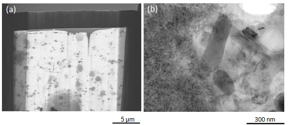
Fig. 1: BF-STEM images of liquid foundation at an accelerating voltage of 30kV(a) and 200kV(b). (a) Instrument: NB5000 FIB-SEM, Cooling temperature: 100K, Magnification: x3,500. (b)Instrument: HD-2700 STEM, Cooling temperature: 100K, Magnification: x50,000. |
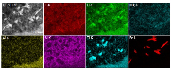
Fig. 2: EDX maps of liquid foundation using the 200kV STEM with the cryo transfer specimen holder. dispersoid dispersantInstrument: HD-2700, Acquisition Pixel: 256×200, Acquisition time: 30 min, Cooling temperature: 100K. |
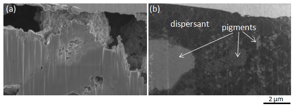
Fig. 3: Cross-section SEM images of liquid foundation. With the mechanical probe at room temperature(a), and with the cryo mechanical probe at 118K(b). Instrument: NB5000 FIB-SEM, Acc. Volt.: 1.5 kV, Magnification: x10,000 |
|